Introduction
Phytophthora
cambivora (Petri) Buisman (1927)
Phytophthora cambivora was first isolated and described as Blepharospora
cambivora, the causal organism of ink
disease of chestnuts in England and the United States, by Petri in 1917
(Petri, 1917). It was further
described as a root rot pathogen
by Buisman
in 1927 in the Netherlands (Buisman,
1927). It is a member of
Waterhouse's group VI species (Stamps et al., 1990). P.
cambivora was at one time regarded
as the sole cause of ink disease of sweet chestnut, but
P. cinnamomi, a more widely distributed species, is known to
be more commonly involved (Waterhouse, 1963; Waterhouse and Waterston,
1966). The pathogen is soilborne and also causes a root rot of other woody plant species (Fig. 1). It can also infect
succulent plants.
Cultural Characteristics
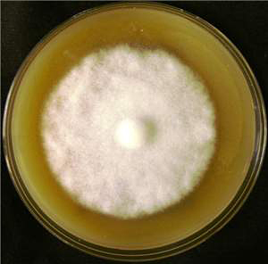
Cultures are uniform, with moderate to profuse
aerial mycelium. The minimum temperature for growth is 2�C, the optimum temperature for growth is 22�24�C, and the maximum temperature for growth
is 32�C (Fig. 2). There is little growth above 30�C.
Reproductive Structures
Asexual
Structures
Sporangiophores:
Sporangiophores are 3�4 µm in diameter and usually unbranched but proliferating within the empty sporangium. Sympodia form in water.
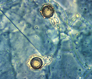
Sporangia:
Sporangia form only in aqueous solutions and
are
broadly ellipsoid or ovoid, usually 40�45 �
55�65 µm (maximum 60 � 85 µm)
(Waterhouse and Waterston, 1966). Sporangia are nonpapillate and
noncaducous, have slight apical thickening, and are not shed. The length�breadth ratio is less than 1.6
(Fig. 3a and b).
Hyphae:
Hyphae are fairly
uniform in diameter and slightly undulate. When old, they are rather broad (7 �m
wide) and thick walled. There are hyphal swellings (in some strains and only on rich
media) with rounded irregular contours that are not usually botryose or coralloid
(Fig. 3c).
Sexual Structures
P. cambivora is normally heterothallic. Sex
organs may be rare or absent in single-strain culture but produced in moderate
abundance in about 10 days when grown with certain strains of
P. nicotianae and
with P. nicotianae
var. parasitica.
Antheridia:
Antheridia are always amphigynous, are often comparatively long, may have one or
two cells, and average 25 µm in diameter (maximum 35 µm).
Oogonia:
Oogonia
are yellow
to brown and average 43 µm in diameter (maximum 62 �m). The oogonial wall
is 2 µm
thick with irregularly disposed bullate protuberances.
Oospores:
Oospores average 36 µm in diameter. Their
walls are 3 µm thick and colorless (Figs. 3d and 4).
Host Range and Distribution
|
Host
|
Common Name
|
Disease
|
Geographical Distribution
|
|
Acer pennsylvanicum
|
Snake-bark maple, whistlewood
|
Root rot
|
United Kingdom
|
|
Castanea
dentata |
American chestnut |
Ink disease |
Europe, United States |
|
Castanea sativa
|
Eurasian chestnut
|
Ink disease
|
France,
Italy,
United Kingdom
|
|
Casuarina equisetifolia
|
Filao, horsetail tree
|
Wilt
|
Mauritius
|
|
Chrysanthemum cinerariaefolium
|
Pyrethrum
|
Wilt
|
India
|
|
Erica spp.
|
Heather
|
Wilt
|
United
States, Spain
|
|
Fagus sylvatica
|
Beech
|
Root rot
|
United Kingdom
|
|
Juglans spp.
|
Walnut
|
Root rot
|
Italy,
Spain
|
|
Malus spp.
|
Apple hybrids
|
Collar and root rot; trunk cankers
|
Japan,
Canada,
United States
|
|
Persea americana
|
Avocado
|
Root rot
|
South Africa
|
|
Pisum sativum
|
Pea
|
Seedling blight
|
Italy
|
|
Prunus spp.
|
Almond, apricot, cherry, European plum, peach
|
Root rot; trunk canker
|
United
States, Australia
|
|
Rhododendron spp.
|
Rhododendron
|
Blight
|
United
States, Denmark
|
|
Rubus idaeus
|
Red raspberry
|
Root rot
|
Scotland
|
|
Senecio spp.
|
Groundsel
|
Root rot
|
United Kingdom
|
|
Ulmus spp.
|
Elm
|
Root rot
|
United Kingdom
|
Symptoms
Ink Disease of Castanea
dentata (Chestnut):
P. cambivora infects the tree near the base of the trunk and on the
larger roots. After infection, the tree generally dies within 2 years. If the
infection spreads rapidly and trees are girdled at the collar, the tree dies within 1 year. If the infection is slower in spreading, leaves and flowers
become smaller during the first year. Inky fluid may discharge from dead and
dying bark at the base of the tree where infection is located.
Root and Crown Rot of Prunus
(Cherry, Peach, Plum,
Almond, and Apricot) and Malus (Apple) Species:
Early stages of infection
are often difficult to detect (Erwin and Ribeiro, 1996). In later stages, leaves
and flowers are smaller; leaves do not grow in terminal shoots. Tree can die
suddenly without observable manifestation of infection. Crown rot and root rot
can be found on the same tree. Crown rot is detected by rotting at the base of
the tree. Root rot is detected by dry, brittle, brown roots. Isolation of the
pathogen is generally the only way to properly diagnose the disease (Wicks and Lee,
1986).
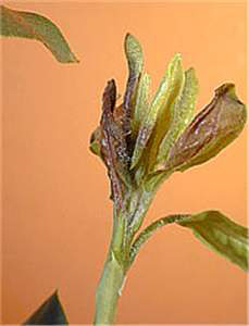
Rhododendron:
The pathogen can infect
the young expanding shoots of rhododendron (Fig. 5).
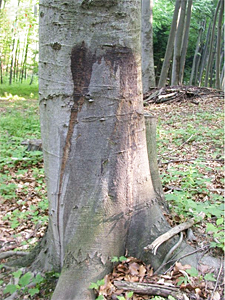
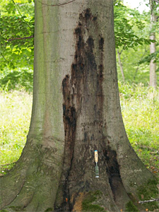
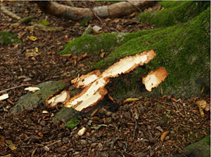
Beech Trees:
The pathogen can infect
the trunk of beech trees and causes lesions that ooze sap (Figs. 6 and
7). Roots can also be infected (Fig. 8).
References
Buisman, C. J. 1927. Root rots caused by
Phycomycetes. Thesis. University of Utrecht. Meded. Phytopathol. Lab. Wille Commelin Scholten 11:7.
Erwin, D. C., and Ribeiro, O. K. 1996.
Phytophthora
Diseases Worldwide. American Phytopathological Society, St. Paul, MN.
Heffer Link, V., Powelson, M. L., and
Johnson, K. B. 2002. Oomycetes. Plant Health Instructor
doi:10.1094/PHI-I-2002-0225-01.
Hwang, J., Warfield, C. Y.,
Parker, K. C., and Benson, D. M. 2006. First report of Phytophthora cambivora
on hybrid rhododendron in North Carolina. Plant Health Progress
doi:10-1094/PHP-2006-0828-01-RS.
Petri, L. 1917. Ricerche sulla morfologia e
biologia della Blepharospora cambivora, parasitica
del castagno. (Research on the morphology and biology of Blepharospora cambivora, parasitica
from chestnut). Atti Regia Accad. Lincei, Rend. Cl. Sci. Fis. Mat. Nat. Ser. 5(26):297-299.
Stamps, D. J., Newhook, F. J., Waterhouse, G.
M., and Hall, G. S. 1990. Revised tabular key to the species of Phytophthora de Bary. Mycol. Pap. 162. CAB International,
Wallingford, United Kingdom;
Commonwealth Mycological Institute, Kew, Surrey, England.
Waterhouse, G.
M. 1963. Key to the species of Phytophthora
de Bary. Mycol. Pap. 92.
CAB International,
Wallingford, United Kingdom;
Commonwealth Mycological Institute, Kew, Surrey, England.
Waterhouse, G. M., and Waterston, J. M. 1966. Phytophthora
cambivora. CMI Descr. Pathog. Fungi Bact. 112:1-2.
Wicks, T. J., and Lee, T. C. 1986. Phytophthora
crown rot in almond trees. Aust. J. Agric. Res. 37:277-287.