Introduction
Phytophthora
alni Brasier
& S. A. Kirk (2004)
Phytophthora alni
was first discovered in 1993 (Brasier et al., 1995) as a Phytophthora
species responsible for alder disease in Alnus trees and was
formally described in 2004 as P. alni by Brasier et al. (2004). The species consists of a range of heteroploid organisms. P. alni subsp. alni is
a tetraploid, while
P. alni subsp. uniformis and P. alni
subsp. multiformis have chromosome numbers between a diploid and a tetraploid. P. alni subsp. alni
was formerly called the standard variant and
P. alni subsp. uniformis
was called
the Swedish variant. P. alni
subsp. multiformis consists of three variants from
Germany,
Denmark, and the
United Kingdom
(Brasier et al., 2004). P. alni subsp.
alni is more aggressive than the other types and is referred to as the
standard variant (Brasier and Kirk, 2001). The pathogen is
believed to be of hybrid origin between
P. cambivora and a
P. fragariae-like species (Brasier et al., 1999). P.
alni subsp. alni may have been
generated on numerous occasions by hybridizations with P.
alni subsp. uniformis and P. alni subsp.
multiformis (Ioos et al., 2006). P. alni subsp. uniformis probably has
P. cambivora as an ancestor, but the origin of P. alni
subsp. multiformis is less clear (Ioos et al., 2006).
These Phytophthora species superficially resembled
P. cambivora in the morphology of its gametangia but differ from
P. cambivora in being self-fertile rather than outcrossing, having a
submerged rather than an aerial colony type, and having different optimum and
upper temperature limits for growth (Brasier et al., 2004).
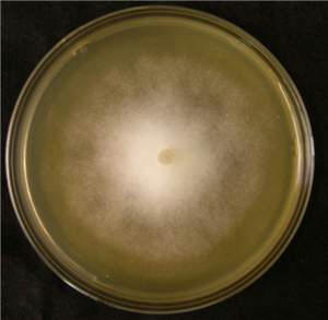
Cultural Characteristics
Cultures have a submerged
colony type with an irregular colony outline (Fig. 1).
P. alni subsp. alni can be grown on carrot agar and has an optimum temperature
for growth of 23¢25░C (Brasier et al., 2004). The maximum temperature for growth is 29░C.
Reproductive Structures
Asexual Structures
Sporangiophores:
Sporangia are borne singly on
long
sporangiophores (Brasier et al., 2004).
Sporangia:
Sporangia are ellipsoid,
nonpapillate, and noncaducous, with a broad
exit pore (Fig. 2). Sporangia are not constricted. The
mean sporangial lengths of
10 isolates range from 48 to 59.8 μm, with an
overall range of 35 to 70 μm. The mean widths range
from 31.3 to 42.8 μm, with an overall range of 27.5 to 50 μm. Mean length¢width ratios
are 1.32¢1.62 (Brasier et al., 2004).
Chlamydospores:
No
chlamydospores have been observed.
Sexual Structures
P. alni
subsp. alni is
homothallic.
Antheridia:
Antheridia predominately
have two cells and are
amphigynous (Fig. 3). The mean lengths of five
isolates range from 23.5 to 27 μm, with an overall range of 20 to 30 μm. The mean widths of antheridia of five isolates range from 18.5 to 19.5 μm,
with an overall range of 15 to 20 μm (Brasier et al., 2004).
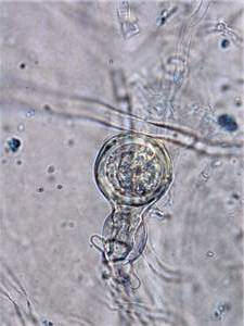
Oogonia:
Some variants of P.
alni subsp. alni have oogonia that are larger and more mature
and have tapered stalks (Fig. 4). The mean diameters of
five isolates range from 42.8 to 50 μm, with an overall range of 37 to 55 μm. Other variants of P. alni subsp. alni have
smaller oogonia with a diameter of 25¢35 μm. Some oogonia
are comma shaped, while others are distorted with beaklike or tubelike
protuberances (Brasier et al., 2004). P. alni
subsp. uniformis forms smooth-walled oogonia, while P. alni
subsp. multiformis and P. alni subsp. alni have
ornamented oogonial walls.
Oospores:
In large oogonia, the mean
oospore diameters of five isolates range from 33.3 to 43.5 μm, with
an overall range of 27.5 to 50 μm (Brasier et al., 2004).
Host Range and Distribution
P. alni
subsp. alni
causes aggressive root and collar rot of Alnus glutinosa and other
Alnus species (Brasier et al., 2004). Alder dieback is now in
11 European countries: Austria,
Belgium, France,
Germany, Hungary,
Ireland, Italy,
Lithuania, Netherlands,
Sweden, and the
United Kingdom (Webber et al., 2004). In the United
Kingdom,
the disease has spread steadily since 1994; by 2003, more than 15% of surveyed
trees were affected or killed (Webber et al., 2004). Alder
dieback is also widespread throughout Austria
(Cech, 2004) and has affected more than one-fourth of the alders along streams
in the Walloon area of Belgium
(Abras, 2005). The pathogen has been found recently in
Alaska (Adams et al., 2008; Trummer et al., 2007).
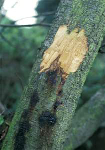
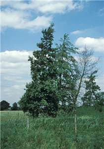
Symptoms
P. alni
subsp. alni forms
a collar rot on alder and dieback. In 1993, scientists in the
United Kingdom determined that the alder
dieback they had observed in recent years was a disease caused by a hybrid
pathogen, P. alni
(Webber et al., 2004) (Figs. 5¢7).
References
Brasier, C. M.,
and Kirk, S. A. 2001. Comparative aggressiveness of standard and variant hybrid
alder Phytophthoras, Phytophthora cambivora, and other Phytophthora species on
the bark of Alnus, Quercus and other woody hosts.
Plant Pathol. 50:218-229.
Brasier, C. M.,
Rose, J., and Gibbs, J. M. 1995. An unusual Phytophthora associated
with widespread alder mortality in Britain. Plant Pathol. 44:999-1007.
Brasier, C. M.,
Cooke, D. L., and Duncan, J. M. 1999. Origin of a new Phytophthora
pathogen through interspecific hybridization. Proc. Natl. Acad. Sci. U.S.A.
96:5878-5883.
Brasier, C. M.,
Kirk, S. A., Delcan, J., and Cooke, D. L. 2004. Phytophthora alni sp.
nov. and its variants: Designation of emerging heteroploid hybrid pathogens
spreading on Alnus trees. Mycol. Res.
108:1172-1184.
Cech, T. L. 2004.
Phytophthora
disease (Phytophthora
alni) of alder - current situation in Austria. Abstr. in: Forstschutz Aktuell Nr.
29¢Abstracts. Bundesforschungs- und Ausbildungszentrum f³r Wald, Naturgefahren
und Landschaft.
http://bfw.ac.at/400/2256.html.
Ioos, R., Andrieux, A., Marcais, B., and Frey, P. 2006. Genetic characterization
of the natural hybrid species Phytophthora alni as inferred from nuclear and
mitochondrial
DNA analyses. Fungal Genet. Biol. 43:511-529.
Trummer, L., Worrall, J., and Adams, G. 2007. Briefing Paper: Phytophthora alni
subsp. uniformis, a first finding in North America.
http://nature.berkeley.edu/comtf/pdf/Alaska_Briefing_Alder_Phytophthora_Nov18.pdf.
Webber, J., Gibbs, J., and Hendry. S. 2004.
Phytophthora
Disease of Alder. Forestry Commission, Edinburgh, United Kingdom.
www.forestresearch.gov.uk/pdf/fcin6.pdf/$FILE/fcin6.pdf.