Introduction
Phytophthora tentaculata Kröber & Marwitz (1993)
Phytophthora tentaculata was first isolated from roots and stalks of the greenhouse-grown ornamental plants of chrysanthemum (Chrysanthemum frutescens hybrid, Chrysanthemum leucanthemum), Delphinium ajacis, and Verbena hybrid and described by Kröber and Marwitz in 1993 (Kröber and Marwitz, 1993). It is closely related to P. multivesiculata according to Kroon et al. (2004). P. tentaculata is a group I Phytophthora species (Stamps et al., 1990).
Cultural Characteristics
The minimum temperature for growth is 7°C, the maximum temperature
for growth is 32°C, and the optimum temperature for growth is 15–25°C.
The growth rate is 3–5 mm per day at optimum temperatures.
Reproductive Structures
Asexual Structures
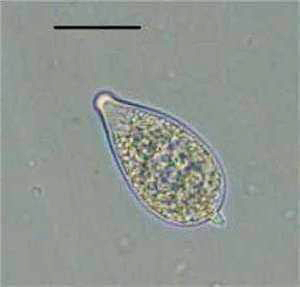
Sporangia:
Sporangia are papillate or
bipapillate and spherical or ovoid to obpyriform. Sporangia are noncaducous but some may
be caducous with a short pedicel.
Sporangia are 13–52 × 10–81 µm (average
27.4 × 35.7 µm) (Fig. 1).
Chlamydospores:
Hyphae:
Hyphal swellings are relatively
small and occur where mycelium
branches. Mycelium appears
arachnoid when grown on carrot agar.
Sexual Structures
P. tentaculata
is homothallic.
Antheridia:
Oogonia:
Oospores:
Host Range
|
Host |
Common Name |
Disease |
Geographical Distribution |
|
Chrysanthemum spp |
Chrysanthemum, Pyrethrum,
Dalmatian flower |
Root and
stem rot |
|
|
Santolina spp |
Santolina |
Root and
stem rot |
|
|
Verbena spp |
Vervain |
Root and
stem rot |
|
Symptoms
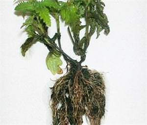
P. tentaculata
causes a root and stem rot of ornamentals.
Heavily infected plants are killed (Kröber and Marwitz, 1993) (Fig.
3).
References
Gallegly, M., and Hong, C. 2008. Phytophthora: Identifying
Species by Morphology and DNA Fingerprints. American Phytopathological Society,
Kröber, H., and Marwitz, R.
1993. Phytophthora tentaculata sp.
nov. und Phytophthora cinnamomi var. parvispora var. nov., zwei neue Pilz von
Zierplantzen in Deutschland (Phytophthora
tentaculata sp. nov. and Phytophthora cinnamomi var. parvispora var. nov., two new fungi from
ornamental plants in
Kroon, L. P. N. M., Bakker, F. T., van den Bosch, G. B. M., Bonants, P.
J. M., and Flier, W. G. 2004. Phylogenetic analysis of Phytophthora
species based on mitochondrial and nuclear DNA sequences. Fungal Genet. Biol.
41:766-782.
Moralejo, E., Puig, M., and Man in 't Veld, W. A. 2004. First report of Phytophthora tentaculata on Verbena sp. in Spain. New Dis. Rep. 9:38.
Stamps, D. J., Waterhouse, G. M., Newhook, F. J., and Hall, G. S. 1990. Revised tabular key to the species of Phytophthora. Mycol. Pap. 162. CAB International, Wallingford, United Kingdom; Commonwealth Mycological Institute, Kew, Surrey, England.