


Introduction
Phytophthora pseudosyringae T. Jung & Delatour (2003)
Phytophthora pseudosyringae is a soilborne pathogen of oak and beech trees. Because of its morphological characteristics, P. pseudosyringae was first identified as P. syringae. RFLP analysis of the internal transcribed spacer (ITS)-DNA sequence data revealed that P. pseudosyringae was more closely related to P. ilicis and P. psychrophila than to P. syringae, and the species name was changed to P. pseudosyringae (Gallegly and Hong, 2008). P. ilicis is the closest relative to P. pseudosyringae. P. pseudosyringae is a group III Phytophthora species (Erwin and Ribeiro, 1996; Waterhouse, 1963) and a member of clade 3 according to Cooke et al. (2000).
Cultural Characteristics
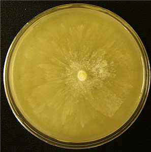
Cultures show a stellate growth pattern on V-8 juice agar and carrot agar (CA) (Fig. 1). On malt extract agar and cornmeal agar (CMA) amended with beta-sitosterol, cultures show a stellate to rosaceous growth pattern. Cultures have an appressed to limited aerial growth on CA and CMA media and limited aerial to fluffy growth on V-8 juice agar. On potato dextrose agar, cultures have appressed and stellate to rosaceous patterns. Not all isolates show the same growth patterns. P. pseudosyringae's temperature range for growth is 5�25�C, and the optimum temperature for growth is 20�C. The maximum temperature for growth is slightly higher for P. pseudosyringae (25�C) than for P. syringae (23�C).
Reproductive Structures
Asexual
Structures
Sporangiophores:
Sporangiophores are
unbranched. Sympodium are simple, lax, or
compact and
sporangia are terminal.
Sporangia:
Chlamydospores:
Hyphae:
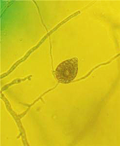
On V-8 juice agar, primary Hyphae are 4�9 μm wide (average 6.2 μm). Hyphae do not branch sympodially. Most isolates produce irregular to coralloid
lateral hyphae
(Jung et al., 2003) (Fig. 6.9). Chains of inflated, spherical to deltoid hyphal swellings
are produced sometimes with radiating hyphae in water culture in isolates from oak and beech and
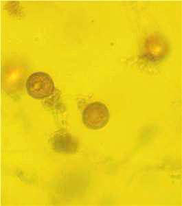
the hyphal swellings average 15.5 μm in diameter (Figs. 2.17 and 2.18 and 4).
Isolates from alder form small numbers of single spherical hyphal
swellings.
Sexual Structures
P. pseudosyringae is homothallic.
Antheridia:
Oogonia:
Oospores:
Host Range
P. pseudosyringae has been found in forest soils in
Symptoms
Symptoms of P. pseudosyringae are similar to the symptoms of
P.
ramorum.
Quercus robur
and Quercus petraea
(Oak):
Fagus sylvatica (Beech):
Alnus glutinosa
(Alder):
Ilex aquifolium
(Holly):
Apple:
References
Cooke, D. E. L., Drenth, A.,
Erwin, D. C., and Ribeiro, O. K. 1996.
Phytophthora
Diseases Worldwide.
Gallegly, M., and Hong, C.
2008. Phytophthora: Identifying Species by Morphology and DNA Fingerprints. American Phytopathological Society,
Jung, T., Nechwatal, J.,
Cooke, D. E., Hartmann, G., Blaschke, M., Osswald, W. F., Duncan, J. M., and
Delatour, C. 2003. Phytophthora
pseudosyringae sp. nov., a new species causing root and collar rot of deciduous tree
species in
Waterhouse,
G. M. 1963. Key to the species of Phytophthora
de Bary. Mycol. Pap. 92. CAB International, Wallingford, United Kingdom; Commonwealth Mycological Institute,