


Introduction
Phytophthora phaseoli Thaxt. (1889)
Phytophthora phaseoli was first described by Thaxter (1889) and amended by
Cultural Characteristics
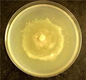
The optimum temperature for growth is 15�20�C, the minimum temperature for growth is 5�C, and the maximum temperature for growth is 25�30�C (Fig. 2). P. phaseoli can be maintained for long periods of time on sterile lima bean seeds in water (Goth and Wester, 1963). Some higher temperature strains have been reported (races E and F).
Reproductive Structures
Asexual
Structures
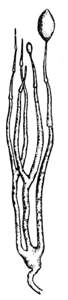
Sporangiophores:
Sporangiophores are sympodially branched and slightly swollen at the base (Fig. 3).
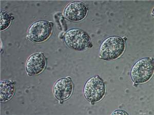
Sporangia:
Sporangia are semipapillate and can be oval or elliptical with a truncate base (Fig. 4). While the size may vary by substrate on which the sporangia are produced (Hyre and Cox, 1953), Thaxter (1890) reports sporangia with dimensions of 20�24 � 3�50 µm. Sporangia are nonproliferating and caducous with short pedicels 5�20 µm long (Fig. 5).
Chlamydospores:
Chlamydospores are not produced.
Hyphal swellings are not produced.
Sexual Structures
P. phaseoli is homothallic.
Antheridia:
Antheridia are amphigynous and shaped like a flattened sphere.
Oogonia:
Oogonia are subspherical and 16.2�35.6 µm in diameter (average 23.7 µm).
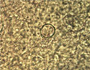
Oospores:
Oospores are produced abundantly on lima bean or host tissue
and are 14.4�26.6 µm in diameter (average 19.2�22.5 �m) (Hyre and Cox, 1953)
(Fig. 6).
Host Range
The pathogen has been found in Africa
(
|
Host |
Common
Name |
Disease |
Geographical
Distribution |
|
Brassica juncea |
Brown mustard |
Seedling
blight |
Philippines (artificially inoculated) |
|
Hevea brasiliensis |
Para rubber |
Seedling blight |
Philippines (artificially inoculated) |
|
Lycopersicon esculentum |
Tomato |
Seedling blight |
Philippines (artificially inoculated) |
|
Phaseolus lunatus, P. limensis, P. vulgaris |
Lima bean, butter bean, common bean |
Downy mildew |
|
|
Raphanus sativus |
Radish |
Seedling
blight |
Philippines (artificially inoculated) |
|
Sandoricum indicum |
Santol |
Seedling
blight |
Philippines (artificially inoculated) |
|
Solanum melongena |
Eggplant |
Seedling blight |
Philippines
(artificially inoculated) |
|
Vigna unguiculata subsp. sesquipedalis |
Asparagus bean |
Seedling blight |
Philippines (artificially inoculated) |
Symptoms
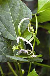
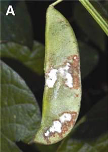
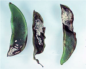
Downy mildew of Phaseolus lunatus (lima bean) is most destructive on pods, but
the pathogen also infects shoots, leaves, and petioles (Fig. 7). Symptoms include early lesions on the
leaves that are irregularly shaped and purplish, and reddish bands often
surround infected areas of the plant (Fig. 8). A white felt of mycelium and
sporangia develops on the pod and eventually progresses to cover the entire
pod (Figs. 9 and 10). Infected
pods eventually shrivel (Fig. 11).
The disease appears most commonly under humid conditions. Disease forecasting systems have been
developed.
References
Blackwell, E. 1949. Terminology in Phytophthora. Mycol. Pap. 30. CAB International, Wallingford, United Kingdom; Commonwealth Mycological Institute, Kew, Surrey, England.
Cline, E. T., Farr,
D. F., and Rossman, A. Y. 2008. A synopsis of Phytophthora with accurate
scientific names, host range, and geographic distribution. Plant Health
Progress doi:10.1094/PHP-2008-0318-01-RS.
Clinton,
G. P. 1906. Downy mildew, Phytophthora
phaseoli Thaxt., of lima beans.
Commonwealth Mycological Institute. 1983. Phytophthora phaseoli. Distrib.
Maps. Plant Dis. 201.
Cooke, D. E. L., Drenth, A.,
Evans, T.
A., Davidson, C. R., Dominiak, J. D., Mulrooney, R. P., Carroll, R. B., and
Antonius, S. H. 2002. Two new races of Phytophthora phaseoli from lima
bean in
Evans, T. A., Mulrooney, R. P., Gregory, N. F., and Kee, E. 2007. Lima bean downy mildew: Impact, etiology, and management strategies for Delaware and the Mid-Atlantic region, U.S. Plant Dis. 91:128-135.
Erwin, D.
C., and Ribeiro, O. K. 1996. Phytophthora Diseases Worldwide. American Phytopathological
Society,
Goth, R.
W., and Wester, R. E. 1963. Culture of Phytophthora
phaseoli on living and sterilized media. Phytopathology 53:233-234.
Hyre, R.
A., and Cox, R. S. 1953. Factors affecting viability and growth of Phytophthora phaseoli. Phytopathology
43:419-425.
Leonian, L. H.
1925. Physiological studies on the genus Phytophthora. Am. J. Bot. 12:444-495.
Stamps, D.
J., Waterhouse, G. M., Newhook, F. J., and Hall, G. S. 1990. Revised tabular
key to the species of Phytophthora.
Mycol. Pap. 162. CAB International, Wallingford, United Kingdom; Commonwealth
Mycological Institute, Kew, Surrey, England.
Thaxter,
R. 1889. A new American Phytophthora.
Bot. Gaz. 14:273-274.
Thaxter,
R. 1890. Report of the mycologist.
Tucker,
C. M. 1931.
Taxonomy of the genus Phytophthora de Bary.
Univ.